

Yuki Keith, Garvan Institute and Tri Phan, Garvan Institute
May 25, 2026
For the past 15 years or so, a class of drugs called immune checkpoint inhibitors have been used to treat melanoma – the most dangerous kind of skin cancer.
For many patients, they produce remarkable results. For others, they do nothing.
We still don’t really know why. But in new research published in the Journal of Experimental Medicine, we observed immune cells called macrophages attacking melanoma cells in real time – which may offer clues about how we can make those therapies work for all patients, not just some.
Tumours, hot and cold
One of us (Yuki) treated patients with melanoma in Japan as a dermatologist. The other (Tri Phan) runs a lab at the Garvan Institute in Sydney, where his team specialises in observing the cells of the immune system in real time.
When Yuki wanted to understand why immune checkpoint inhibitors were failing for many patients, she joined Tri Phan’s lab to continue her research.
The treatment fails in what oncologists call “cold” tumours, where the cancer’s environment actively prevents a kind of immune cell called a T cell attacking it. One of our lab’s aims is trying to work out how to make the tumours “hot”, allowing T cells to penetrate and destroy the cancer cells.
Our new findings suggest a different kind of immune cell, called macrophages, may hold the key.


The housekeepers we’ve been ignoring
In 1908, Russian zoologist Ilya Mechnikov was awarded a Nobel Prize for the discovery of phagocytosis (“cell eating”) in the immune system, which is carried out by cells he called macrophages (from the Greek for “big eaters”).
These cells engulf and clear away the debris caused by tissue damage and cell death. They are often regarded as the body’s silent, no-fuss housekeepers.
However, their role in cancer has often been overlooked. Unlike other immune cells that move through the blood and patrol the whole body, macrophages are “tissue-resident” and stay in one place.

Earlier studies of the role of macrophages in cancer assumed these housekeepers were all the same. But when we looked closely in the skin, it became clear that there were many different kinds of macrophages living in different layers.
One particular kind of macrophages (recognised by a protein called CD169) lives in a deeper part of the skin, called the hypodermis.
We found that these macrophages arranged themselves around the edges of a melanoma tumour, as if they were trying to wall it off. When we depleted the macrophages, the melanomas grew bigger, suggesting they were constraining the growth of the tumours.
Watching cancer cells being eaten alive
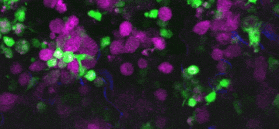
To understand what these CD169-positive macrophages were actually doing, we used an advanced imaging technique called intravital two-photon microscopy. This allows us to watch biological processes unfold in living tissue in real time.
What we saw was surprising: the macrophages were “nibbling” and actively engulfing live melanoma cells. While we had seen macrophages eat dead cells in our lab before, we had never seen them eat a live melanoma cell in a model organism.
What was even more surprising was that this immune attack was happening without the need for T cells, or antibodies made by another kind of immune cell called B cells – the immune players most commonly credited with fighting cancer.
We also confirmed this is not something that just happens in the lab. Our colleagues at the Melanoma Institute Australia analysed samples from human melanoma patients and found similar populations of CD169-expressing macrophages on the edges of the tumour, suggesting they may play a similar protective role there.

Calling in the cavalry – implications for therapies
Macrophages don’t just clear away debris. They can also alert the immune system to danger. After they have digested the debris, they can display it like a biological “red flag” to direct T cells to find and kill the cancer cells.
What makes a macrophage decide whether to silently dispose of debris without alerting the immune system, or wave the red flags to activate the immune system, is still unclear. Because the CD169-expressing macrophages are strategically positioned around the tumours, we suspect they may hold the key.
Macrophages are widespread in most solid tumours – including glioblastoma, breast cancer and many others. This is an army already in place waiting to be mobilised.
Our next step is to understand precisely how these macrophages eat live cancer cells and how they can communicate the danger to T cells, so we can harness this population with new treatments.

Click & Grow Garden
Sleek and simple indoor smart gardens that help you grow fresh herbs and vegetables with ease—no experience needed. Shop Click & Grow and support The-14 through our affiliate link.
Visit StoreYuki Keith, Postdoctoral Researcher, Immunology, Garvan Institute and Tri Phan, Program Director – Precision Immunology / Laboratory Head, Garvan Institute
Subscribe to our newsletter.
This article is republished from The Conversation under a Creative Commons license. Read the original article.
